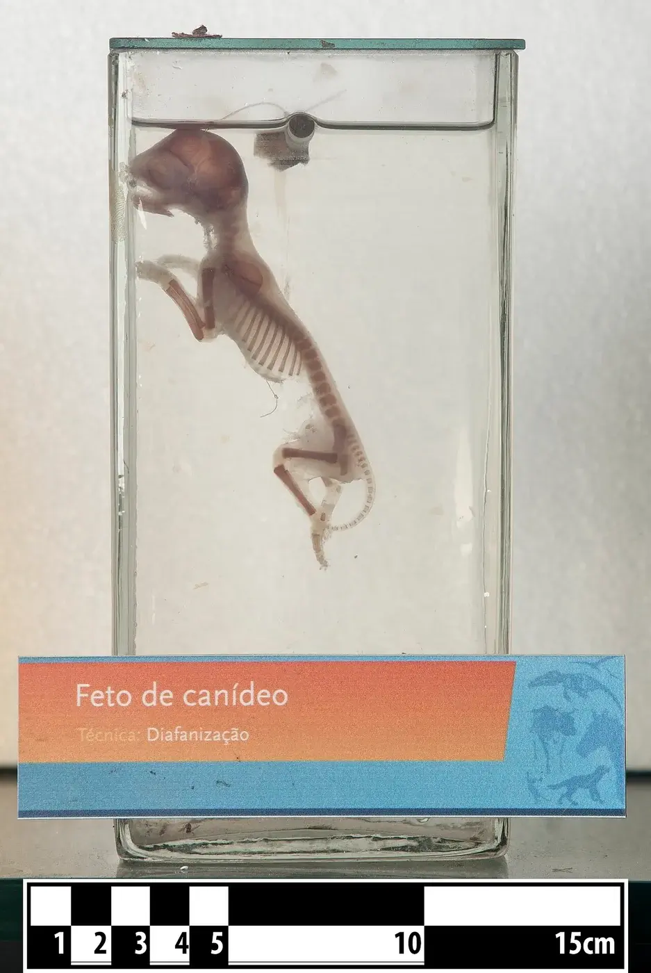
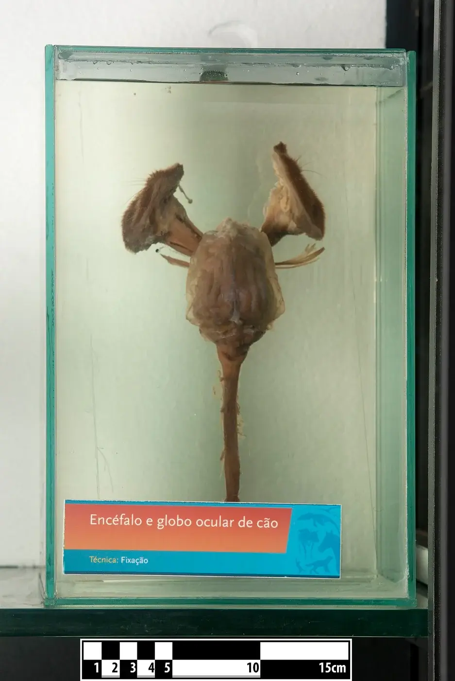
Dog Night Vision KevKan · CC BY-SA 4.0 · Web size optimized; source linked for the original.
A dog’s eyes let it track a ball in dim light or notice small movements that people miss. The lens and retina work as a team: the lens bends light, and the retina catches it with specialized cells. Scientific papers describe the lens as a clear structure that passes light unaltered to the retina, and the retina as a layered tissue containing rods for low-light vision and cones for daylight and color.
The Lens Passes Light to the Retina
The lens sits behind the cornea. It is optically clear and has no blood vessels. Its layers run from an anterior capsule through the cortex and nucleus to a thin posterior capsule. Its only job is to let light and images reach the retina without distortion.
As dogs age the lens nucleus can become denser. This change is called nuclear sclerosis and is considered a normal part of aging rather than a cataract. Cataracts are opacities that interfere with light passage; nuclear sclerosis is different.
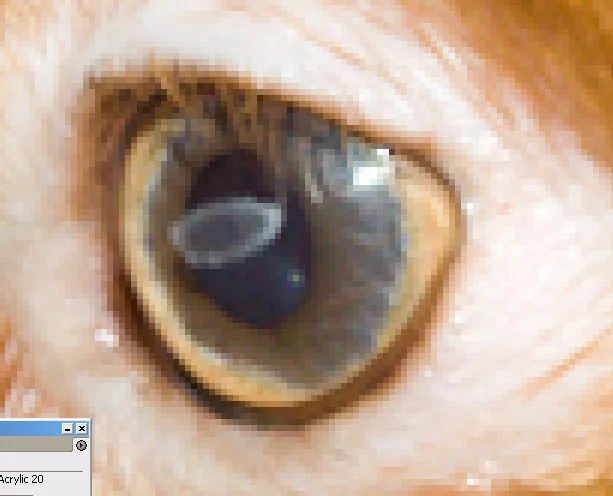
Close-up of a dog's eye Ttrimm · CC BY-SA 3.0. Web size optimized; original linked.
Retinal Layers and Photoreceptors
The retina lines the back of the eye and has ten layers whose fine structure resembles that of other mammals, with some minor differences. Photoreceptors sit in the outer part of the retina. Rods handle low light and motion detection; cones handle brighter conditions and color.
Rods and cones are highly polarized cells. Their inner and outer segments differ in shape and length between the two cell types. In dogs, rods outnumber cones, which supports vision in dim settings.
Development of the Retina After Birth
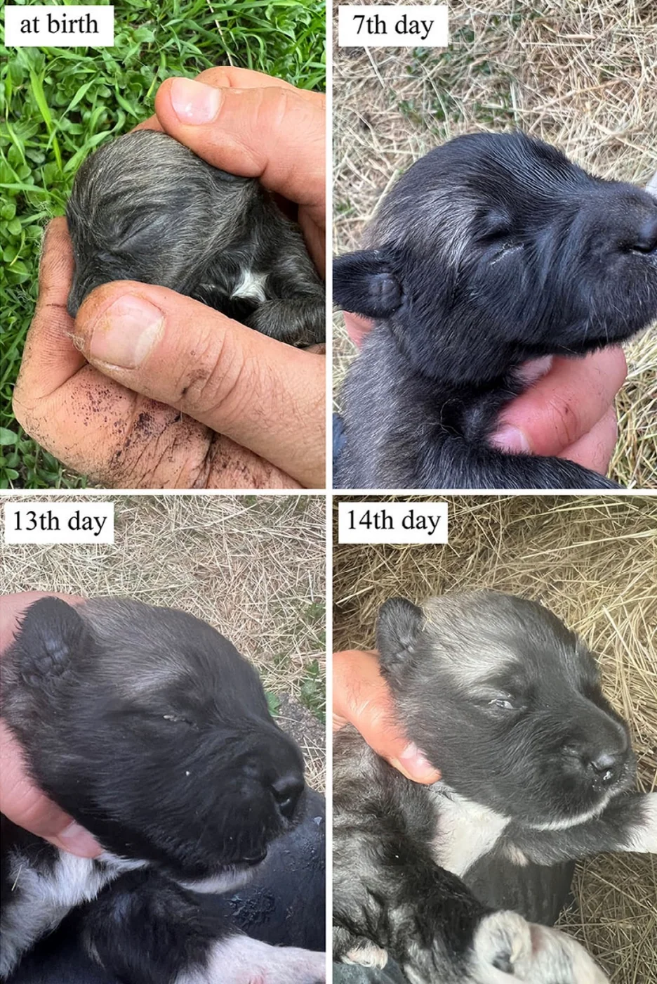
Retinal maturation continues after puppies are born. The process moves from the central retina outward. By about 16 days after birth, slender rod and cone segments appear in the central retina. These segments reach the peripheral retina roughly 35 days after birth.
This timetable comes from one specific study of developing canine eyes. The study does not describe lens development or how the maturing lens and retina interact.
The supplied papers focus on either the lens or the retina but do not detail how the two structures coordinate in a living dog. The lens is described only as the structure that must remain clear for light to reach the retina. Retinal studies examine the ultrastructure of photoreceptors and the timing of their maturation.
No source supplied here explains color vision, field of view, motion detection in breeds, or the tapetum lucidum. Those topics cannot be covered because they lack supporting excerpts.
Why Owners Might Notice These Structures
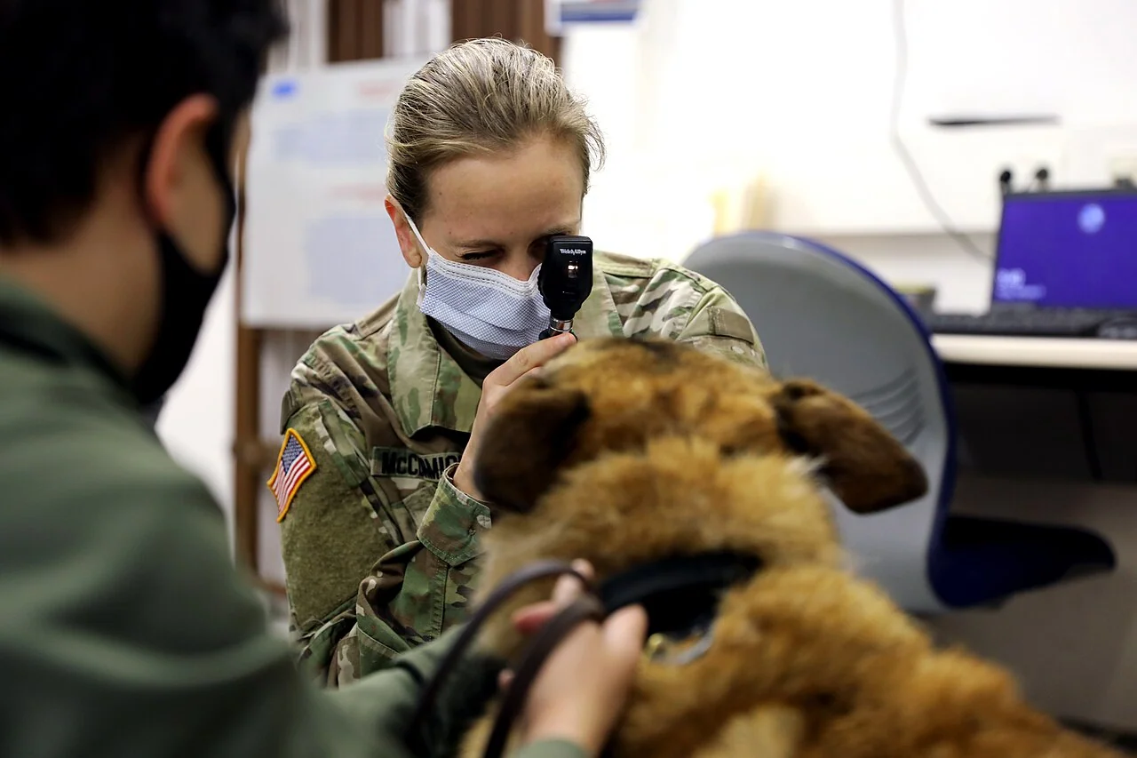
When a dog’s lens clouds or its retina is affected, light may not reach the photoreceptors properly. Routine veterinary exams can observe the lens and the visible parts of the retina. The Merck Veterinary Manual notes that distinguishing cataracts from normal aging helps owners understand what they see.
The research papers are laboratory studies of eye tissue. They do not give advice on pet care, breeding, or treatment.
Veterinary officer conducting a routine eye exam on a military working dog, demonstrating how owners and veterinarians observe lens and retinal health U.S. Army RHCE by Russell Toof · Public domain. Web size optimized; original linked.
General education, not individual veterinary advice. Ask a veterinarian about your dog’s health and care.
AI-assisted original writing. This feature was revised against source material and checked for unsupported claims. Follow the references below for evidence and its limits.
Research shows canine cognition follows a clear arc of early gains, midlife peak, and later gentle decline, offering owners practical ways to support their pets at every age.
Protected command centers in the brain and spinal cord team up with far-reaching nerves that connect to muscles, skin, and organs, letting dogs react to scents, sounds, and sudden surprises.

 Reference image