Practical Limits of fMRI When Imaging Awake Dog Brains
Scanning awake canines offers a window into their neural responses, yet motion, small samples, and uncertain haemodynamic timing require careful reading of every result.
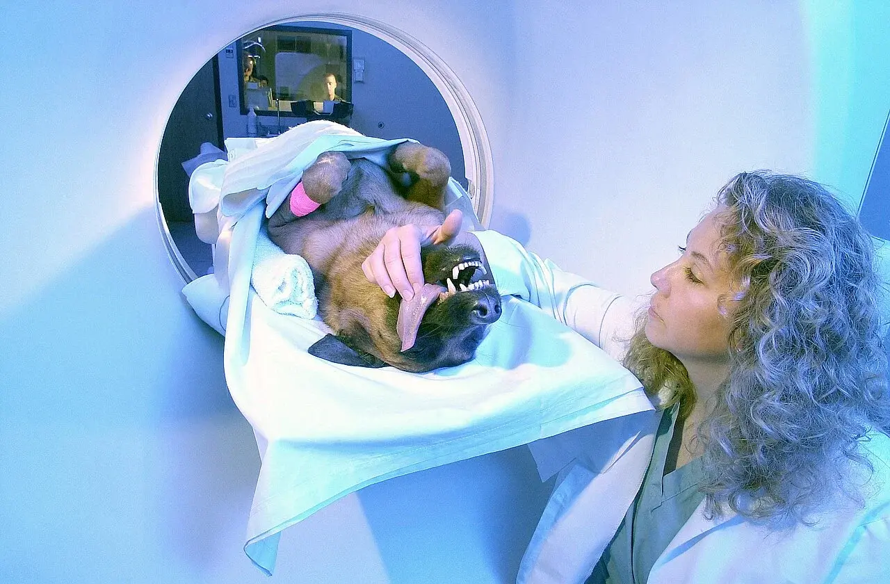
Dog lying motionless with bandaged head in a human knee coil inside the MRI scanner bore (2020 training study) Karl S, Boch M, Virányi Z, Lamm C, Huber L. · CC BY 4.0 · Web size optimized; source linked for the original.
Functional magnetic resonance imaging (fMRI) has opened new possibilities for exploring how dogs process the world around them without the confounding effects of anesthesia. Researchers can now watch brain activity while dogs respond to voices, smells, or rewards. Yet the very features that make these studies appealing—working with awake, behaving animals—also introduce substantial technical and statistical obstacles. Evidence from multiple studies shows that data quality can suffer from subject movement, uncertain timing of blood-flow responses, and the small number of dogs that can be trained for scanner work. These limitations do not invalidate the research, but they do call for measured interpretation rather than sweeping conclusions about canine minds. The following sections examine the main challenges, what researchers are doing to address them, and what readers should keep in mind when encountering claims based on awake-dog fMRI.
Why Awake Scanning Matters
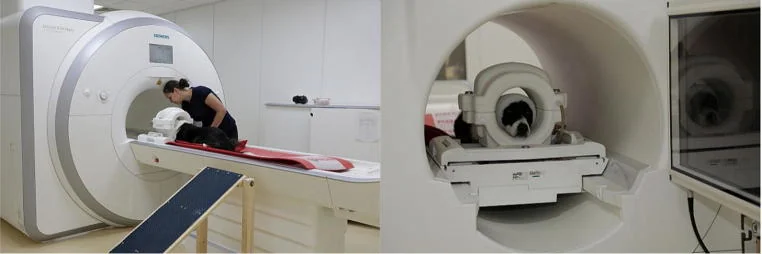
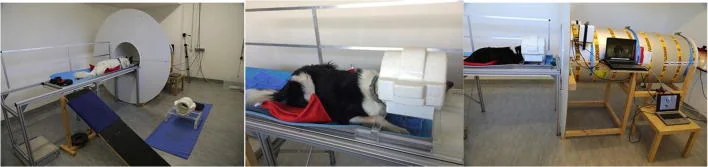
Anesthesia changes blood flow, breathing patterns, and neural signaling in ways that can mask the very cognitive processes scientists hope to study. For questions about reward, emotion, or social cognition, an unconscious animal simply cannot provide relevant data. Training dogs to lie still inside a noisy scanner removes the need for sedation and lets researchers observe brain responses during real behavioral tasks. This advance has produced intriguing glimpses—such as activation in reward-related areas when dogs anticipate a treat. Yet the practical cost of that advance is high: only a limited number of dogs tolerate the procedure, and even well-trained ones still move more than human volunteers.
The trade-off is therefore clear. Awake imaging preserves ecological validity at the expense of data cleanliness. Each successful scan represents hours of patient training, but the resulting datasets are noisier and smaller than typical human neuroimaging studies. Editorial perspective suggests this is a worthwhile trade, provided the community remains transparent about the resulting uncertainty. Without such candor, popular headlines risk overstating what a handful of tail-wagged scans can truly tell us about “what dogs are thinking.”
Even after extensive training, dogs move more than humans during scans. Head motion creates artifacts that can mimic or hide genuine neural signals. Researchers address this by discarding volumes that exceed motion thresholds, adding motion parameters as nuisance regressors, and sometimes using real-time motion tracking. In early work, roughly 38–42 percent of scan volumes were excluded because of movement, meaning a large fraction of expensive scanner time yields no usable data.
The problem is not trivial. Residual motion after censoring can still bias statistical maps, especially when sample sizes are modest. Light-hearted observation: imagine trying to solve a jigsaw puzzle while the dog in the next room is wagging its tail hard enough to rattle the table. The humor fades when one realises that each discarded volume reduces statistical power and increases the chance that findings are driven by a few unusually compliant animals rather than a reproducible biological effect. Future hardware improvements, such as better parallel imaging, may ease the burden, but motion remains the dominant practical limitation today.
The Haemodynamic Response Function Problem
Human fMRI analysis typically assumes a standard haemodynamic response function (HRF) that peaks roughly six seconds after neural activity. Canine brains differ in size, blood-vessel density, and neurophysiology, so the actual timing and shape of their HRF are unlikely to match the human template exactly. Using the wrong model reduces detection power, inflates apparent effect sizes, and can increase false-positive rates. One line of work has therefore developed dog-specific HRF templates that improve sensitivity without sacrificing specificity.
Even with tailored models, uncertainty lingers because the precise HRF in dogs has not been mapped across breeds, ages, or arousal states. This gap matters: minor mismatches in timing can turn a statistically significant result into noise or vice versa. Editorial analysis: the field has moved beyond simply borrowing human pipelines, yet readers should still treat peak coordinates and activation strengths with caution. A useful question for any new paper is whether the authors verified their HRF choice against dog physiology or simply adopted the newest published template.
Most awake-dog fMRI studies scan between 10 and 15 animals—far fewer than the dozens or hundreds typical in human cognitive neuroscience. Small samples magnify the impact of individual differences and outliers. A single highly responsive dog can drive a group-level finding that fails to replicate. Compounding this, the number of trials per dog is often limited by fatigue and attention span, further reducing power to detect subtle effects.
Underpowered studies risk both missing real effects and publishing inflated ones. When only a handful of dogs contribute usable data, the probability that an observed activation reflects a genuine, generalizable phenomenon drops. One constructive implication is that meta-analyses and multi-site collaborations will become increasingly important. Until those exist, consumers of this literature should ask: how many dogs actually entered the final analysis, and what fraction of collected data survived motion scrubbing? Transparent reporting on these points helps separate solid findings from statistical mirages.
Reading the Literature Responsibly
Given the constraints outlined above, how should non-specialists approach claims about canine emotion or decision-making derived from fMRI? A healthy skepticism is warranted but need not become outright dismissal. Look for studies that report motion metrics, use dog-optimized HRF models, and acknowledge the modest sample. Consistency across independent laboratories adds confidence, especially when different training protocols converge on similar brain regions.
Reading questions worth keeping in mind include: Does the paper quantify data loss due to motion? Are the statistical maps corrected for multiple comparisons across the whole brain? Is the claimed activation spatially reliable, or does it shift several millimeters between animals? On the lighter side, remember that even the most rigorous scan cannot tell you why your dog stole the cheese; it can only hint at which circuits lit up while the theft was contemplated. That distinction between correlation and everyday explanation is worth preserving. Ultimately, awake-dog fMRI is a promising tool whose limitations are not fatal but instructive. They remind us that understanding another species’ mind requires converging evidence from behavior, physiology, and imaging—none of which is perfect on its own.
AI-assisted original writing. This feature was revised against source material and checked for unsupported claims. Follow the references below for evidence and its limits.
Archaeological finds from select European sites offer glimpses into possible relationships between people and dogs, while underscoring the challenges of interpreting ancient deposits.
Owner-led projects offer scale and real-world glimpses into canine minds, yet differences in tester identity and uncontrolled home settings can introduce systematic errors that large samples do not automatically cancel out.
Penn State researchers once used MRI scans and computer simulations to map airflow inside a dog’s nasal passages, offering clues for designing better chemical sensors.
 Photograph